These are good CT images to review, along with self-test questions and answers.
Pulmonary Embolus (figure 4):

|
Explain the CT findings.
This is a CT pulmonary angiogram, which uses contrast to visualize the veins during venous phase. Contrast within the veins is seen as areas of high attenuation. Thrombus within the vessel is seen as a 'filling defect' (a low attenuation area as there is no contrast there). If seen in an axial plane through the vessel it has the appearance of a polo mint.
|
Colonic Polyp (figure 12):
|
Why is it important to remove bowel polyps?
Polyps have the potential to develop into colorectal carcinoma, especially in those suffering from FAP.
|
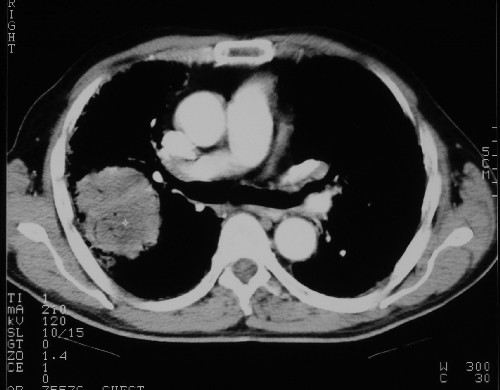
Bronchial Carcinoma (figure 14, 15):
|
What are the subtypes of bronchial carcinoma?
- Squamous cell carcinoma,
- adenocarcinoma,
- large cell
- small cell (oat cell)
The subtype dictates subsequent management.
Those suspected of having a bronchial carcinoma require CT chest and abdomen (for staging) and if appropriate bronchoscopy with biopsy specimens or washings.
|
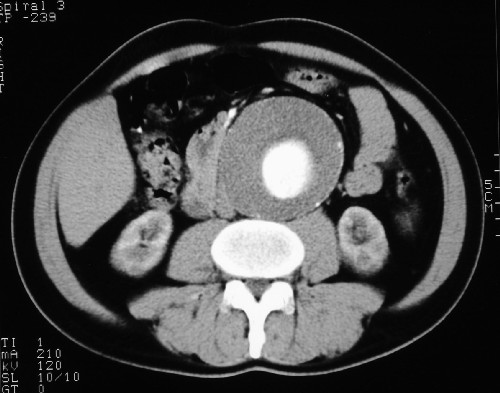
Abdominal Aortic Aneurysm (figure 17):
|
What size of aneurysm would be considered for repair?
Those aneurysms reaching 5.5cm are candidates for elective surgery. Most procedures are still by open repair, with trials ongoing of endovascular stenting - especially in those with poor co-morbid health.
|
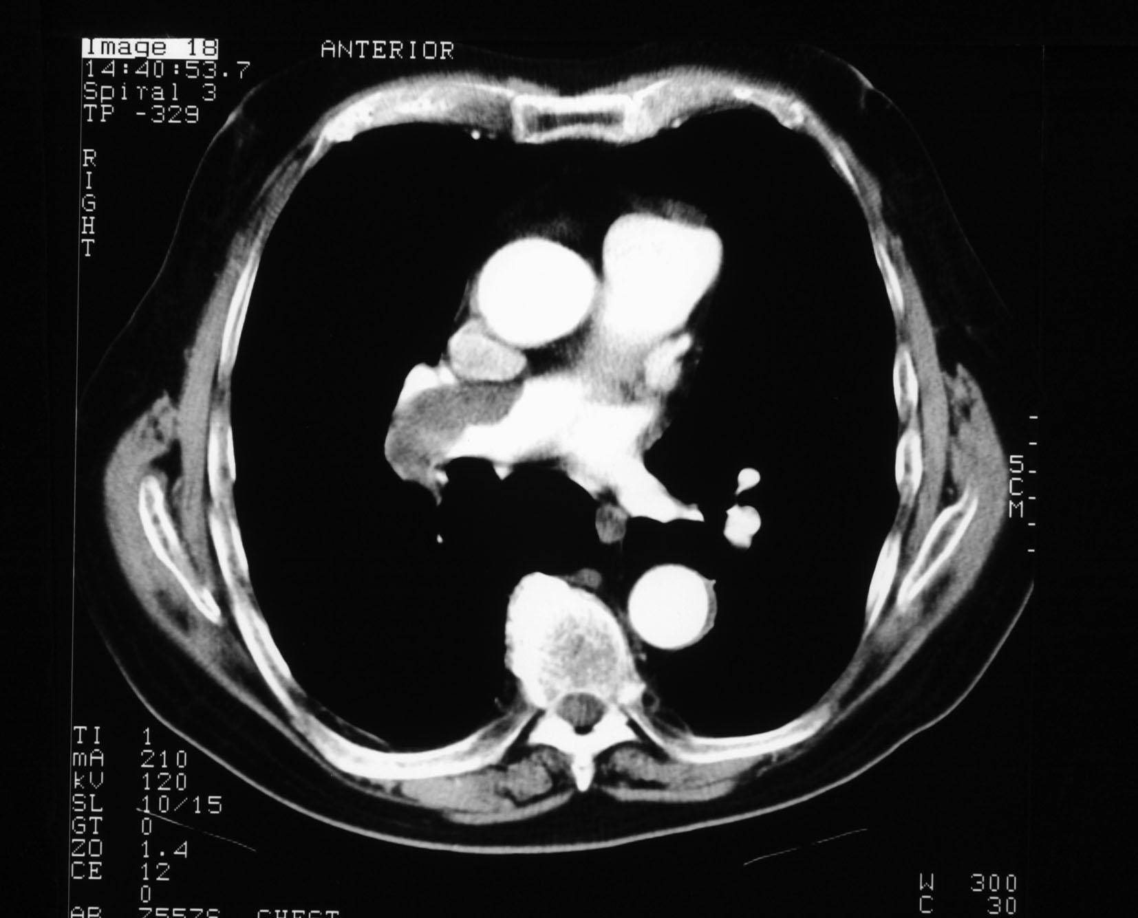
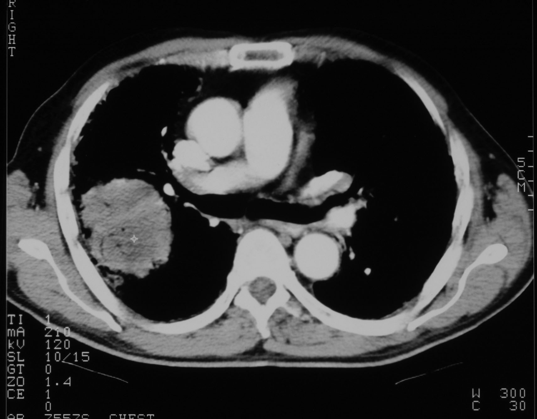
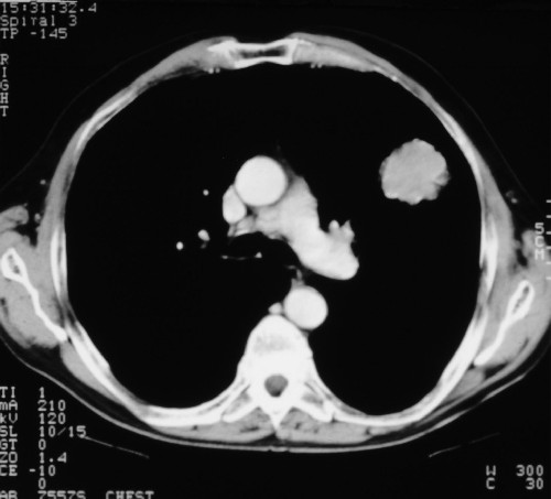
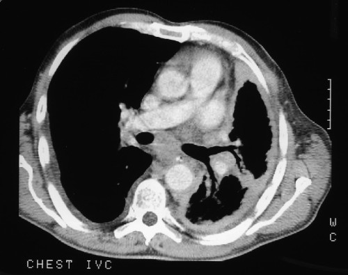
Malignant Mesothelioma (figure 18):
|
What chest disease can occur following exposure to asbestos?
- Malignant mesothelioma
- Pleural plaques
- Asbestosis
- Bronchial carcinoma
|