| Medical Finals contents: Welcome Finals advice Written exams Clinical revision X-Rays Mock final OSCE's |  |
Medical Finals contents: Monthly quiz PDA's Links Credits Your comments Who are we? |
| Medical Finals contents: Welcome Finals advice Written exams Clinical revision X-Rays Mock final OSCE's | |
Medical Finals contents: Monthly quiz PDA's Links Credits Your comments Who are we? |
Question 1 | ||||||||||||||||||
|
This 35 year-old man was admitted to hospital with reduced loss of consciousness.
A CT head (non-contrast) was performed: 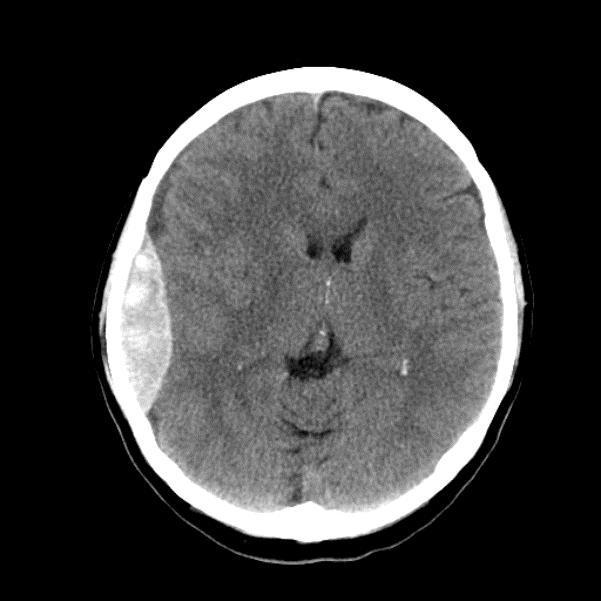
Which of the following intracranial abnormalities is shown? | ||||||||||||||||||
| ||||||||||||||||||
|
Answer: (d) Extra-dural haematoma
Extra-dural and sub-dural haematoma are extra-axial collections.
|
Question 2 | |||||
|
This 44 year-old man presented to his GP with visual problems.
MRI brain was performed: 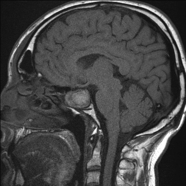
Which of the following defects is most likely to be found on examination? | |||||
| |||||
|
Answer: (b) Bilateral temporal hemianopia
The MRI shows a pituitary tumour. Pituitary adenomas can be divided into marcoadenomas and microadenomas. A macroadenoma is greater than 10mm in size and is the more common type. The tumours can be further classified into functioning and non-functioning depending on whether they exhibit endocrine activity. The most common functional tumour is a prolactinoma. The normal pituitary lies within the pituitary fossa, but when it enlarges compresses on the superiorly situated optic chiasm causing a bilateral temporal hemianopia (bitemporal hemianopia). See more on the visual pathway and defects at: http://www.unmc.edu/Physiology/Mann/pix_7/vis_field.gif |
Question 3 | |||||
A 55 year-old man presents with left sensori-neural hearing loss. His MRI scan shows a cerebellopontine angle mass:
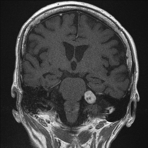
Which of the following is the most common cerebello-pontine angle mass? | |||||
| |||||
|
Answer: (d) Vestibular schwannoma (acoustic neuroma)
Masses at the cerebellopontine angle may present clinically with a cerebellopontine angle syndrome. This is caused by a space occupying lesion at the junction of the cerebellum and the pons, which includes important structures such as the Vth, VIIth and VIIIth cranial nerves, Signs of CPA syndrome include:
The commonest cause (accounting for approximately 80% of masses) is a vestibular schwannoma (acoustic neuroma). These are usually benign tumours of the vestibular portion of the VIIIth cranial nerve. They are associated with neurofibromatosis Type 2. Neurofibromatosis Type 2 (remember 2!)
|
Question 4 | |||||
This 29 year-old man has been admitted to the neurology ward on several occasions during the past 3 years.
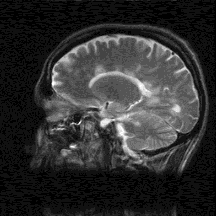
Which of the following is the mode of inheritance of this condition? | |||||
| |||||
|
Answer: (c) Sporadic
This is a classical appearance of multiple sclerosis on MRI. MRI is the premier imaging modality for visualizing the inflammatory demyelinating plaques which are a feature of the disease. Plaques may occur in the cerebral hemispheres, cerebellum, brainstem, and spinal cord, classically in a periventricular distribution (see image). However, multiple sclerosis is a clinical diagnosis, supported by imaging and CSF analysis findings. It is a sporadic disease process. |
Question 5 | |||||
|
A 27 year-old lady attends early pregnancy assessment clinic with her partner and is distressed to learn that she has had a miscarriage, with a 7 week non-viable pregnancy. This is her third in 4 years. On reviewing her notes you note a raised anti-cardiolipin antibody titre in her laboratory results section.
What other clinical problem would fit with the diagnosis of anti-phospholipid syndrome? | |||||
| |||||
|
Answer: (d) Deep venous thrombosis
Anti-phospholipid syndrome (APS) is a condition characterized by recurrent venous or arterial thrombosis and/or fetal losses in conjunction with persistently elevated levels of antibodies directed against membrane phospholipids typically anti-cardiolipin antibody. Several autoimmune diseases are associated with APS, the commonest being systemic lupus erythromatosis. |