| Medical Finals contents: Welcome Finals advice Written exams Clinical revision X-Rays Mock final OSCE's |  |
Medical Finals contents: Monthly quiz PDA's Links Credits Your comments Who are we? |
| Medical Finals contents: Welcome Finals advice Written exams Clinical revision X-Rays Mock final OSCE's | |
Medical Finals contents: Monthly quiz PDA's Links Credits Your comments Who are we? |
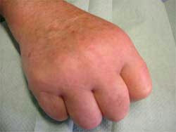
John is a 50 year old car mechanic. He presents to his GP with an 8 week history of pains in his hands, elbows, shoulders, knees and feet. He has remarked that his joints have become quite 'swollen'. They are stiff in the morning time and take more than 30 minutes to 'warm up'. On examination you notice that his MCP joints are symmetrically swollen, red and tender. Clinically the GP diagnoses early Rheumatoid Arthritis (RA).
 What would be the recommended course of action that the GP should take? |
| (a) Refer to a rheumatologist only when his pains become quite severe. |
| (b) GP should commence Methotrexate |
| (c) Routine referral to a rheumatologist |
| (d) Urgent referral to a rheumatologist - THE CORRECT ANSWER |
| (e) X-ray his hands |
|
(d) Urgent referral to a rheumatologist
Rheumatoid Arthritis (RA) is an inflammatory arthritis that has a prevalence of 1%. It is a severely debilitating disease; 50% of patients with RA will have considerable physical limitations & disability. Typically patients present with symmetrical joint pains mainly affecting the hands/feet, early morning stiffness (i.e. symmetrical stiffness that takes greater than 30mins to improve after exercising / movement). Clinically the patient has synovitis in their MCP's joints (i.e. they are swollen, red, warm and tender on the MCP 'squeeze test'). 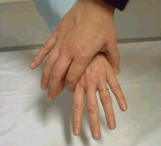 MCP Squeeze test If a GP observes clinical joint swelling they are 40 times as likely to diagnosis RA. To aid the diagnosis of RA, the symptoms have to be present greater than 6 weeks. Viral arthritis (which in its early stages can be indistinguishable from early RA) usually settles down before 6 weeks from onset. Early diagnosis and aggressive treatment with Disease Modifying Anti Rheumatic Drugs (DMARDs) is paramount. Such early aggressive treatment has been proven to improve outcomes in patients with RA. Generally it is inadvisable for a GP to prescribe a DMARD. They usually need initiated by a Rheumatologist. In the early stages of RA there usually is no radiological evidence of inflammation or erosions and a hand xray may be reported as 'normal'. However an MRI scan may pick up early signs of inflammatory arthritis such as periarticular bone oedema. 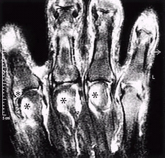 * = MRI evidence of periarticular bone oedema An MRI scan of a patients hand 4 weeks after developing symptoms of early RA. Periarticular bone oedema is apparent in the second, third and fourth MCP joints. |
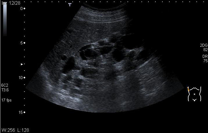
The following patient with hypertension had an ultrasound of the abdomen due to an elevated urea and creatinine.
 What is the likelihood of his sister having the same condition? |
| (a) 25% |
| (b) 0% |
| (c) 50% - THE CORRECT ANSWER |
| (d) 100% |
| (e) 75% |
|
(c) 50%
This patient has adult polycystic kidney disease (APKD). It is inherited by an autosomal dominant transmission. Given that this patient has APKD it would indicate one of his parents carries the gene. It is not sex linked. His sister is therefore equally likely to inherit the condition. APKD is characterized by a multitude of cysts within the kidneys (see ultrasound). It may present with hypertension, haematuria or loin pain. A significant proportion will develop renal failure requiring renal replacement therapy. Cysts may be found in other organs, including the liver and pancreas. There is an association with cerebral berry aneurysm and so subarachnoid haemorrhage. |
|
A 21 year-old female student notices an enlarging mass in her neck. On examination it lies in the upper 1/3 of the anterior triangle. It is fluctuant in nature.
What is the most likely diagnosis? |
| (a) Cystic hygroma |
| (b) Branchial cyst - THE CORRECT ANSWER |
| (c) Thyroid cyst |
| (d) Lymph node |
| (e) Carotid body tumour |
|
(b) Branchial cyst
Neck masses may be divided clinically into those situated in the anterior and posterior triangles of the neck. The anterior triangle is bounded by the midline anteriorly, sternocleidomastoid muscle posteriorly and superiorly by the inferior margin of the mandible. The posterior triangle is bound by the sternocleidomastoid muscle medially, the clavicle inferiorly and laterally by the anterior border of the trapezius muscle. The clinical description indicates the mass is in the anterior triangle.
A branchial cyst typically lies in the anterior triangle at the upper 1/3 of the sternocleiodmastoid muscle. It is a remnant of the branchial complex and classically presents between the ages of 15-30 years of age. It is fluctuant being a cyst. Fine Needle Aspiration (FNA) is the gold standard for diagnosis of any neck lump and should be undertaken to confirm diagnosis. |
|
A 71 year-old man complains to his GP of bony aches and pains. He is otherwise well. Bone profile sent.
AST, ALT, GGT are normal. What is the most likely diagnosis? |
| (a) Prostatic carcinoma with bony metastases |
| (b) Osteoporosis |
| (c) Primary sclerosing Cholangitis |
| (d) Paget's disease - THE CORRECT ANSWER |
| (e) Osteomalacia |
|
(d) Paget's disease
Three of the diagnoses offered in the answer are possible. The following may all cause an isolated raised ALP:
Given the absence of any other disease and normal liver function indices PSC is unlikely. The prostate specific antigen is normal and no mention is made of urinary symptoms, however Paget's disease is common and in a proportion with manifest as generalized or localized bone pain. |
| When assessing an antero-posterior (AP) chest x-ray (CXR) which structure(s) should be commented on with care? |
| (a) Trachea |
| (b) Pulmonary arteries |
| (c) Heart - THE CORRECT ANSWER |
| (d) Lung Apices |
| (e) Breast shadows |
|
(c) Heart
The most commonly requested imaging investigation and hence the most commonly requested radiological based exam question is to present/interpret a chest radiograph. Cardio-respiratory disease accounts for a significant proportion of acute medical admissions and consequently findings on CXR often dictate medical management. The film should be fully illuminated on a light box, with as little finger contact as possible, not least in the heat of an exam, to avoid sticky fingerprints! As with any film if there are two of anything, always use these for comparison. Initially comment on the film specifics. The exact details of projection (AP, PA, Semi-erect), the type of film (CXR, AXR), and the date the film was taken. Be carefully commenting on the size of the heart on an antero-posterior (AP) projection due to magnification. It may be part of a series and thus other films are needed for accurate comparison and comment. The patient details are added to the introductory statement, chiefly the name and age and perhaps on occasion the location (eg, cardiac ICU). Technical assessment follows, with assessment of rotation, inspiration and penetration (RIP). There should be equidistance between the spinous processes and the medial end of the clavicle. Change in this implies rotation and thus alteration in the dimensions of other structures. Ten posterior or greater than five anterior ribs should be visible at the level of the midclavicular line. The thoracic vertebrae should be clearly seen at the mid thoracic spine if properly penetrated. One is now in the position to look for anatomical pathology in a systematic fashion, viewing the following areas; heart, lungs and pleura, bones and soft tissues, review areas. There are 3 key aspects of the cardiac evaluation; heart size (cardiothoracic ratio), heart borders and the pulmonary vascularity. The latter reflects the pulmonary wedge pressure which increases in cardiac failure. To review pulmonary structures; check the trachea is central, the size, shape and level of the hilia, the lung fields, comparing one side with the other. There are 4 key points to review with regard to the diaphragm; its level, shape, the presence of any subdiaphragmatic gas and whether the costophrenic angles are blunted. Finally, view the pleura for any thickening or evidence of a pneumothorax. With regard to the bones and soft tissues; check the ribs, clavicle and humerus for fractures and metastatic involvement. You may note any other generalised bone diseases too. Surgical emphysema and soft tissue masses may be noted. Breast shadows are important in women - the loss of one suggesting previous mastectomy for breast carcinoma. Don't forget to comment on any artefactual findings; pacemakers, sternotomy wires, coronary stents being just a few. |