| Medical Finals contents: Welcome Finals advice Written exams Clinical revision X-Rays Mock final OSCE's |  |
Medical Finals contents: Monthly quiz PDA's Links Credits Your comments Who are we? |
| Medical Finals contents: Welcome Finals advice Written exams Clinical revision X-Rays Mock final OSCE's | |
Medical Finals contents: Monthly quiz PDA's Links Credits Your comments Who are we? |
Question 1 | ||||||||||||||||||
|
A 65 year old man presents to his GP with fatigue and loss of appetite.
On examination: jaundice; palpable non-tender mass in the right upper quadrant. The results of his liver function tests are shown:
Urinalyis showed the presence of bilirubin. Urobilinogen was undetectable. Alpha feto-protein was normal. What is the most likely diagnosis? | ||||||||||||||||||
| ||||||||||||||||||
|
Answer: (a) Pancreatic Carcinoma
Jaundice is defined as a yellow discoloration of the skin, is is typically detectable when the serum bilirubin level is over 50µmol/L. The causes of jaundice can be classified under three broad headings: pre-hepatic, hepatic, and post-hepatic. Liver function tests are one of the most useful investigations used to give an indication of the underlying cause. In pre-hepatic jaundice:
In hepatic jaundice:
In post-hepatic jaundice:
In this case, there is a moderate rise in the liver enzyme ALT but a marked rise in the ALP which is suggestive of an obstructive/post-hepatic picture. The presence of bilirubin but absence of urobilinogen in the urine also supports this picture. Hepatocellular carcinoma may cause an obstructive picture, but the normal alpha feto protein suggests this is less likely. Therefore the most likely diagnosis here is pancreatic carcinoma. The diagnosis of the cause of jaundice cannot be made on the basis of the liver function tests alone. Other investigations such as ultrasonography, MRCP, ERCP, and CT should be considered. |
Question 2 | |||||
|
A 60 year old woman attends her GP with a lump in her right breast. The lump was noticed when she checked her breast after a fall on her chest. There is no nipple discharge. On examination there is a palpable fixed mass approximately 4cm x 3cm in the upper outer quadrant, with a smooth border. Mammogram showed a dense opacity. Histopathology ruled out breast cancer.
Which of the following is the most likely diagnosis? | |||||
| |||||
|
Answer: (b) Fat necrosis
The most likely diagnosis is fat necrosis. Fat necrosis typically occurs in elderly women with large breasts, or after injury to the chest, for example following a car accident when the seatbelt has squeezed the breast. This clinically presents as a firm painless lump, although there may be some redness or bruising. The only way to exclude breast cancer is FNAC. Fibroademomas, or "breast mice", typically affect young women between the ages of 15 and 25 years. They are usually well circumscribed, firm, smooth mobile lumps. Investigation with triple assessment (clinical breast exam, breast histopathology and radiology) can diagnose the condition and exclude breast cancer. Breast cysts and duct ectasia are disorders of involution that occur most often in older women. Breast cysts present as smooth discrete masses that can be painful. They are seen on mammography as "halos" and on ultrasound. Most cysts are asymptomatic and do not require treatment. Symptomatic cysts can be aspirated, but recurrence, or blood stained fluid on aspiration is an indication for surgical excision. Duct ectasia typically presents with cheesy discharge and "slit like" nipple retraction. Treatment is only indicated if the discharge is worrisome or if the patient wants their nipple everted. Duct papillomas are benign neoplasms that are very common. They usually present with blood-stained nipple discharge. Duct papillomas show little malignant potential, but treatment involves removal of the discharging duct, and allows exclusion breast cancer. |
Question 3 | |||||
A 35 year old woman complains to her GP of a facial rash, with purple discolouration of the eyelids and a swollen eye. She has also noticed some non-pruritic bluish/red nodules over her knuckles.
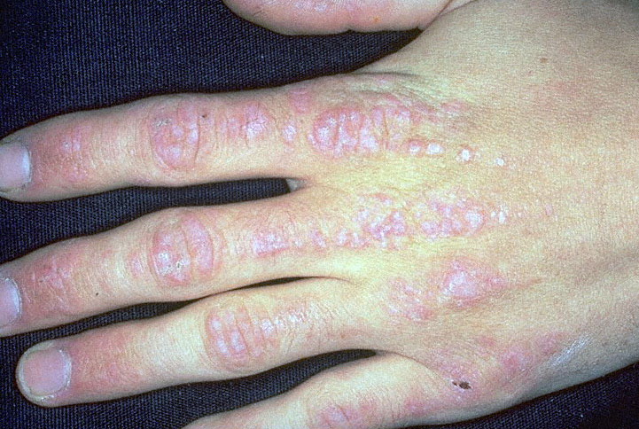
Dermatomyositis picture What is the most likely diagnosis? | |||||
| |||||
|
Answer: (b) Dermatomyositis
This is the classic presentation of dermatomyositis. Dermatomyositis and polymyositis are rare disorders that involve inflammation of striated muscle causing weakness of proximal muscles. When the skin is involved, it is called dermatomyositis. Clinical features include facial erythema, a purple "heliotrope" rash on the eyelids and periorbital area. Bluish/red nodules typically occur on the extensor surfaces and over the knuckles (Gottron's papules). Diagnosis of the condition involves:
|
Question 4 | |||||
|
A 74 year old man presents to A&E with a suspected fracture of his femur. He has experienced aching bone pain for approximately 6 months. He is also hard of hearing.
He has a raised serum alkaline phosphatase and normal serum calcium. PSA is normal. His x-ray shows an expansile sclerotic process. What is the most likely diagnosis? | |||||
| |||||
|
Answer: (c) Paget's Disease
Paget's disease is a common disease in middle-aged and older people. Accelerated turnover of bone, with increased osteoblastic and osteoclastic activity results in disorganised bone architecture which leads to changes in the mechanical properties of the bone. Paget's disease may cause expansion of the membrane bones of the skull, with compression the acoustic nerve and deafness. In addition, pagetic bone is typically brittle, and fracture can occur with minimal or no trauma.
|
Question 5 | |||||
A 25 year old woman notices a red, painful rash on her shins.
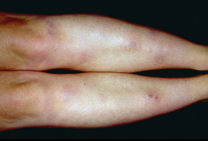
Erythema Nodosum picture Which of the following would you consider as a cause? | |||||
| |||||
|
Answer: (e) All of the above
The history is suggestive of erythema nodosum, which presents as painful nodules, usually on the extensor aspect of the lower limbs. It is most common in young adults, especially females. It is due to inflammation of the dermis and the subcutaneous layer (panniculitis). There may be associated arthralgia, malaise and fever. Common causes of erythema nodosum include:
Other less common causes include:
All patients presenting with erythema nodosum require a CXR given the differential diagnoses.
|