| Medical Finals contents: Welcome Finals advice Written exams Clinical revision X-Rays Mock final OSCE's |  |
Medical Finals contents: Monthly quiz PDA's Links Credits Your comments Who are we? |
| Medical Finals contents: Welcome Finals advice Written exams Clinical revision X-Rays Mock final OSCE's | |
Medical Finals contents: Monthly quiz PDA's Links Credits Your comments Who are we? |
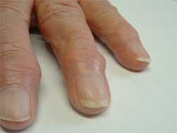
Mary is a 68 year old retired cleaner. She presents to her GP with pains in her 'hand joints'. She is finding simple tasks like opening buttons increasingly difficult. Her pains tend to get worse as the day progresses. Her hands do get stiff particularly after use. She has never noticed her hands to be swollen but has noticed little 'swellings on the ends of her fingers'. Clinically you do not detect any signs of synovitis.
  What would be recommended first line pharmacological treatment for this patient? |
| (a) High dose NSAIDs |
| (b) Paracetamol - THE CORRECT ANSWER |
| (c) Methotraxate |
| (d) Topical NSAIDs |
| (e) Sulfasalazine |
|
(b) Paracetamol
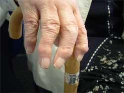
This patient has nodal osteoarthritis (OA). This is a condition of synovial joints characterized by focal areas of damage to the articular cartilage and remodelling of underlying bone. OA affects up to 20% of the population who are greater than 60yrs of age. The main symptoms of OA include pain and immobility. In the photos graphs you can see classical Heberden's nodes in her DIP joints. Similar nodes at the PIP joints are termed Bouchard's nodes. The CMC joint in the hand is also commonly involved in nodal OA. Interestingly this lady has OA of the hip - hence the walking stick. In most cases paracetamol is an effective and relatively safe treatment for patients with mild-moderate OA pain. NSAIDs have been proven to be effective in reducing OA pain but they do carry risks such as gastric ulceration; adverse renal effects and precipitating relapse of heart failure. Up to 1.5% of NSAID users per year develop serious upper GI complications such as a major bleed or perforation. More patients die of NSAID related upper GI bleeds, ulceration and perforations per year in the UK than RTA related deaths. There is conflicting evidence that topical NSAIDS have long term benefit, if any in OA. DMARDS have no place in the treatment of OA. |
|
A 20 year old man with a history of type I diabetes is admitted with pyrexia, drowsiness, and fast deep breathing. You suspect diabetic ketoacidosis (DKA).
What is the initial treatment priority? |
| (a) IV drip of 5% dextrose solution |
| (b) IV drip of 0.9% Saline - THE CORRECT ANSWER |
| (c) 10 units of subcutaneous short acting insulin |
| (d) Insulin via infusion pump |
| (e) Potassium supplementation |
|
(b) IV drip of 0.9% Saline
Patients with DKA, tend to be significantly dehydrated due to osmotic diuresis, and reduced fluid intake. In adults this is usually at least 3.5 to 7 litres. A treatment plan for DKA would typically be:
|
|
During a finals short case, you palpate a significantly enlarged liver and spleen.
What is the commonest cause of large hepatosplenomegaly in UK/Ireland? |
| (a) Zinc deficiency |
| (b) Haematological disease - THE CORRECT ANSWER |
| (c) Chronic alcohol abuse |
| (d) Infectious mononucleosis |
| (e) Malaria |
|
(b) Haematological disease
Hepatosplenomegaly is enlargement of both the spleen and liver. Causes include: (i) Haematological disease - Myeloproliferative disease, Leukaemia, Lymphoma, Pernicious anaemia, Sickle cell anaemia, Thalassaemia. (ii) Acute Infection - Acute viral hepatitis, Infectious mononucleosis, Cytomegalovirus. (iii) Chronic liver disease (including Chronic alcohol abuse, Chronic active hepatitis, NASH, Amyloidosis, Acromegaly, Systemic lupus erythematosus), with portal hypertension. In the early stages, cirrhosis can cause liver to enlarge, and indeed alcoholic hepatitis can produce tender hepatomegaly, but later as more scar tissue replaces normal tissue, the liver shrinks. |
|
As on-call PRHO (foundation year 1) you are called to see a 52 year old post-op patient who is hypotensive (BP=80/60). You bleep your SHO who advises a fluid challenge.
Which of the following fluid regimes would be most appropriate? |
| (a) 1 litre 5% dextrose over 4 hours |
| (b) 500 ml Colloid (Gelofusion or Voluven) over 60 minutes |
| (c) 1 litre Normal saline over 4 hours |
| (d) 250ml Normal Saline over 2 minutes - THE CORRECT ANSWER |
| (e) 1 unit of reheated fresh frozen plasma over 30 minutes |
|
(d) 250ml Normal Saline over 2 minutes
Often colloid (eg. Gelofusion or Voluven) is recommended as it stays in blood vessels longer - but crystalloid (eg. Normal Saline) can be used. The important thing is to give a small volume rapidly - this will not run through a drip counter (as drip-counter max rate is typical 999ml/hour) - whereas 250ml Normal Saline over 2 minutes = 7500ml/hour (although some suggest using a longer time of 10 minutes). A wide bore (16 or 14 gauge venflon) is needed. Measure the blood pressure before, during and 5 minues after this bolus. If the pulse falls and BP rises this indicates the patient is hypovolaemic (absolute or relative) eg. from blood loss or sepsis, so more fluid should be given. It is also useful to note the duration of any BP rise. (For more info see: Surgical Tutor - Fluid balance and: Student BMJ - Fluid challenge) Causes of hypotension: (use mnemonic CPR):
|
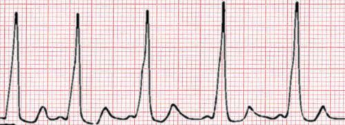
You are called to see a patient in A+E, and are shown the following rhythm strip and ECG:
 What does this show? |
| (a) Ventricular tachycardia (VT) |
| (b) Atrial Fibrillation (AF) |
| (c) First degree heart block |
| (d) Agonal rhythm |
| (e) Wolf-Parkinson-White (WPW) - THE CORRECT ANSWER |
|
(e) Wolf-Parkinson-White (WPW)
The ECG has P-waves so is not Atrial Fibrillation (AF). Is not First degree heart block as that would have long PR interval. QRS's are narrow so is not VF nor VT. Is not an Agonal rhythm which would be extreme ventricular bradiacardia, with wide flattened QRS. It is Wolf-Parkinson-White (WPW), as has shortened PR interval (less than 3 squares < 120ms) and delta waves (slurred upstroke to the QRS indicating pre-excitation), as shown in diagram below. This delta wave is the classical feature. The QRS may be broad, and there can be secondary secondary ST and T wave changes. WPW is due to an accessory pathway between atria and ventricles. Patients present with SVT which may be due to AV re-entry tachycardia, pre-excited AF, or pre-excited atrial flutter. In symptomatic patients, the ideal treatment is to use radio frequency catheter ablation to destroy the accessory pathway. 
WPW showing shortened PR and delta wave. A good illustration of normal, and Wolff-Parkinson-White Syndrome accessory path-way. (For more info see ECG links on Links page on www.medicalfinals.co.uk.) |