| Medical Finals contents: Welcome Finals advice Written exams Clinical revision X-Rays Mock final OSCE's |  |
Medical Finals contents: Monthly quiz PDA's Links Credits Your comments Who are we? |
| Medical Finals contents: Welcome Finals advice Written exams Clinical revision X-Rays Mock final OSCE's | |
Medical Finals contents: Monthly quiz PDA's Links Credits Your comments Who are we? |
Question 1 | |||||
A 64 year old man is being treated for his lung abscess (see chest x-ray).
 He develops a headache and nausea. His family report he is increasingly irritable. His investigations show:
What is the most likely complication to cause his symptoms? | |||||
| |||||
|
Answer: (c) SIADH (Syndrome of Inappropriate ADH)
Syndrome of inappropriate ADH (SIADH) results from excess secretion of ADH (vasopressin). ADH causes water re-absorption in the distal tubule, and excess action of ADH causes an inappropriately high urine osmolality, with a low plasma osmolality and low serum sodium. There are multiple causes (see box below):
Diabetes incipidus (DI) presents with polyuria and polydipsia. The pattern of plasma and urine osmolality seen in DI is the reverse of SIADH. A water deprivation test with synthetic ADH is used to distinguish between cranial and nephrogenic DI. This may also be used to distinguish CDI and NDI from Psychogenic Polydipsia. Cushing's syndrome rarely presents acutely and is due to high levels of steroids, which may be exogenous (e.g. long term prednisolone), or endogenous. Dehydration may present with headache and irritability, but would cause a normal-high plasma osmolality, concentrated urine (high osmolality) and a high plasma urea. |
Question 2 | |||||
|
One of the antibiotics considered in this man's treatment plan has side effects listed as liver enzyme induction, nausea, and red discolouration of bodily secretions.
Which is the most likely drug to cause this? | |||||
| |||||
Answer: (b) Rifampicin
In patients prescribed rifampicin it is important to warn patients about the red-secretions, as this can stain contact lenses, and cause undue worry. Isoniazid may cause peripheral neuropathy, so prophylactic Pyridoxine (vit B6) is usually given. Isoniazid also may cause hepatitis. Ethambutol is also used to treat TB, it causes the important side effect of retrobulbar neuritis, which presents with colour blindness and a central scotoma. Visual field testing is performed before commencement of ethambutol for baseline status. |
Question 3 | |||||
|
Q3. During a teaching ward round your clinical supervisor takes you to evaluate a chest x-ray of a child. On inspection you notice the heart is boot shaped and the lungs are oligaemic.
What is the most likely congenital cardiac defect? | |||||
| |||||
|
Answer: (b) Tetralogy of fallot
Tetralogy of Fallot is an example of congenital cyanotic heart disease.
The initial investigation of congenital heart disease includes a chest x-ray, ECG and echocardiography.
|
Question 4 | |||||
This 68 year-old man attends his GP complaining of a short history of a red, hot, swollen great right toe (see image below). He has a history of hypertension for which he is on treatment.
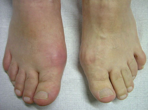 The clinical diagnosis given by the GP is acute gout. Which is the most likely drug to have precipitated acute gout? | |||||
| |||||
|
Answer: (e) Bendroflumethiazide
The differential diagnosis for an acute, red, hot swollen joint is:
Gout is an inflammatory arthritis due to the deposition of urate crystals in the joint. It is associated with a raised serum uric acid but is not required for the diagnosis to be made. There are a number of causes of gout, including prescribed medications.
Treatment is divided into those medications for acute gout and those for chronic gout:
|
Question 5 | |||||
This 58 year old patient presents to his GP with the following condition.
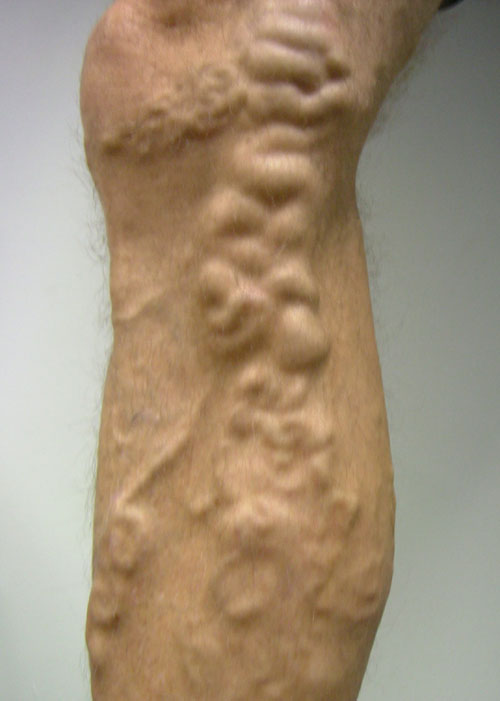 Which of the following is not a recognised complication of this condition? | |||||
| |||||
|
Answer: (e) Deep venous thrombosis
Varicose veins are dilated, tortuous superficial veins in the lower limb. It is a common condition which most often prompts clinical advice for cosmetic reasons, although a small proportion of patients have symptoms or complications. Complications of varicose veins include:
There is no evidence from studies that having varicose veins leads to a DVT. |