| Medical Finals contents: Welcome Finals advice Written exams Clinical revision X-Rays Mock final OSCE's |  |
Medical Finals contents: Monthly quiz PDA's Links Credits Your comments Who are we? |
| Medical Finals contents: Welcome Finals advice Written exams Clinical revision X-Rays Mock final OSCE's | |
Medical Finals contents: Monthly quiz PDA's Links Credits Your comments Who are we? |
Question 1 | |||||
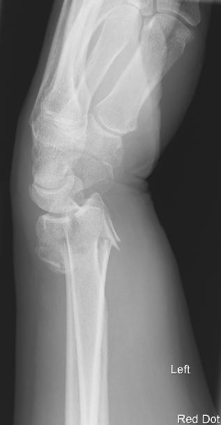
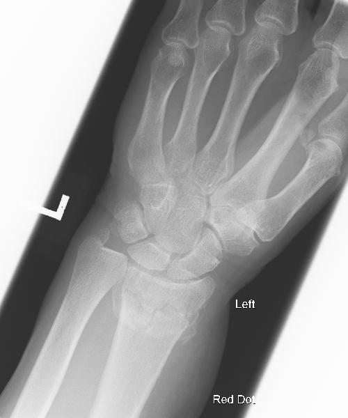
Which of the following is correct regarding a Colle's fracture?
  | |||||
|
Answer: (b) the fracture occurs within 2.5cm of the wrist joint
Colle's fractures represent over 90% of distal radius fractures. They display the classic 'dinner fork deformity' on x-ray. The definition of a Colle's fracture is:
|
Question 2 | |||||
|
A four week old male infant presents with difficulty feeding. Mum is worried that he vomits (forcefully) after his feeds, and this is becoming more frequent. In addition, he is constantly hungry despite feeding well and has failed to gain any weight. Stools are normal.
On examination a palpable 'olive' mass is felt in the right upper quadrant. What is the most likely diagnosis? | |||||
| |||||
|
Answer: (c) pyloric stenosis
All of the above diagnoses should be considered in an infant presenting with vomiting or regurgitation. However, the history above is consistent with pyloric stenosis. Pyloric stenosis typically presents between 2 and 8 weeks of age. It is four times more common in males, particularly first borns, and may be associated with a family history. Classic features are:
Examination of the abdomen often reveals an 'olive' mass in the RUQ. Clinical diagnosis may be made by using a test feed the baby is given a milk feed, and the abdomen examined: visible gastric peristalsis is seen as a wave moving left to right across the abdomen. If the abdomen is distended, this may be difficult to appreciate. Ultrasound is the primary imaging modality to confirm the diagnosis. Treatment is initially with correction of fluid and electrolytes abnormalities followed by pyloromyotomy. |
Question 3 | |||||
|
Which of the following is TRUE regarding a Monteggia fracture?
A Monteggia fracture is best described as a fracture of the: | |||||
| |||||
|
Answer: (a) ulna with dislocation of the head of the radius
The mechanism of injury for a Monteggia fracture is usually following a direct blow to the arm OR forced pronation (e.g. a fall on an outstretched arm accompanied by rotation of the trunk) The definition of a Monteggia fracture is "a fracture of the ulna with dislocation of the head of the radius". |
Question 4 | |||||
|
A 6 year old boy presents to A&E with a short history of abdominal and joint pain. He is otherwise well and apyrexic. You notice a non-blanching purpuric rash on his legs.
What diagnosis would be your primary concern? | |||||
| |||||
|
Answer: (b) Henoch-Schonlein purpura
Henoch-Schonlein purpura (HSP) usually occurs between the ages of 3 and 10 years. It is more common in males, has a peak in winter, and is often preceded by a URTI. The characteristic features include:
Over 80% of patients with HSP have either microscopic or macroscopic haematuria or mild proteinuria at presentation. The disease is usually self-limiting, but nephrotic syndrome is a rare complication. |
Question 5 | |||||
|
A four month old infant presents with 'abnormal jerky movements'. Mum describes repeated violent flexor spasms of the head, trunk and limbs followed by extension of the arms. It is associated with intermittent loss of consciousness. The spasms most commonly occur in the morning, shortly after wakening.
The following EEG was obtained: 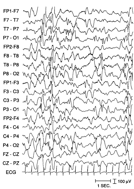
(Reprinted with permission from eMedicine.com.) What diagnosis would you consider? | |||||
| |||||
|
Answer: (a) West syndrome
The EEG above shows hypsarrhythmia a chaotic background of high-voltage dysrhythmic slow-wave activity with multi-focal spike and wave discharges. This EEG is characteristic of 'West syndrome' or infantile spasms. West syndrome usually presents between the age of four and six months, with males and female equally affected. The spasms described in the case above are characteristic of the disease. Other features of the disease include impaired social interaction and developmental regression. Prognosis is poor with most infants showing loss of skills and later learning disability or epilepsy. For more details see: eMedicine.com - Infantile Spasm (West Syndrome). |