| Medical Finals contents: Welcome Finals advice Written exams Clinical revision X-Rays Mock final OSCE's |  |
Medical Finals contents: Monthly quiz PDA's Links Credits Your comments Who are we? |
| Medical Finals contents: Welcome Finals advice Written exams Clinical revision X-Rays Mock final OSCE's | |
Medical Finals contents: Monthly quiz PDA's Links Credits Your comments Who are we? |
Question 1 | |||||
A 72 year old lady presents to the ophthalmology department after sudden blindness in her right eye. The following fundal appearance is noted.
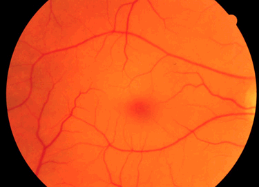 (Click on image for sharper view. The click your brower's BACK button to return to this page) Which of the following most accurately describes the fundus? | |||||
| |||||
|
Answer: (b) Central retinal artery occlusion
This demonstrates a cherry red spot and is diagnostic of a central retinal artery occlusion. |
Question 2 | |||||
|
You are the ophthalmology SHO.
What is your immediate management? | |||||
| |||||
|
Answer: (d) Ocular massage
Immediate management is ocular massage to try and dislodge the embolus. |
Question 3 | |||||
|
On further questioning this lady alludes to having had severe headaches prior to the onset of sudden blindness.
What investigation would you like to perform? | |||||
| |||||
|
Answer: (e) ESR
An ESR would be most helpful in this context as the clinical history is highly suspicious of temporal arteritis. |
Question 4 | |||||
|
You have the result of the above investigation to hand.
What is your next step in her management? | |||||
| |||||
|
Answer: (c) High dose steroids
She must be immediately commenced on high dose immunosuppression with oral prednisolone. A temporal artery biopsy is advised to confirm the diagnosis. Treatment with steroids in the interim will not alter the biopsy result significantly. |
Question 5 | |||||
An x-ray of the wrist is shown below.
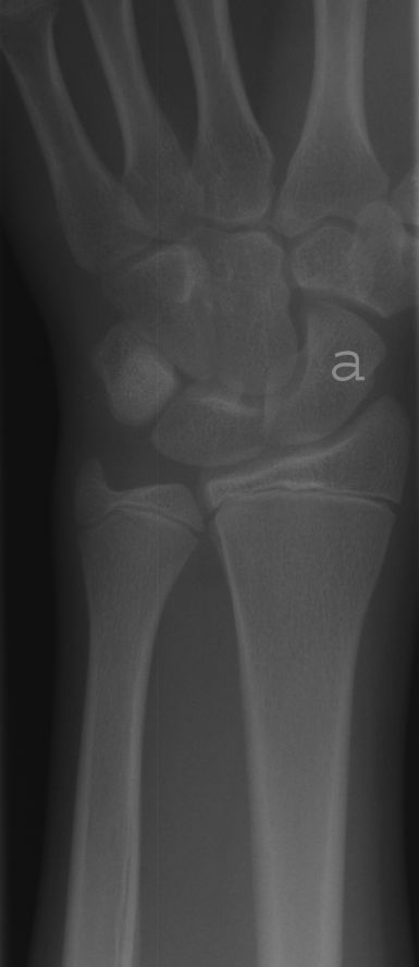 (Click on image for enlarged view. The click your brower's BACK button to return to this page) Which of the carpal bones is labelled 'a'? | |||||
| |||||
Answer: (d) Scaphoid
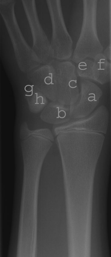 (Click on image for enlarged view. The click your brower's BACK button to return to this page) There are 2 rows of carpal bones in the wrist - the proximal and distal. Distal Row: d = Hamate
Proximal Row: g = Pisiform
|